Javascript is required to use this site, but javascript is not enabled in your browser.
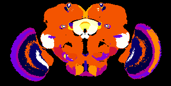
Find out about the braincode project.